What an MRI Shows in a Corgi Back
It's an utterly heartbreaking experience when your usually lively Corgi suddenly starts showing signs of back pain, weakness, or even paralysis. One minute they're zooming around the yard, the next they're struggling to stand, climb stairs, or even just move comfortably. The fear and uncertainty can be overwhelming, leaving you desperate for answers and a clear path forward. This guide is here to walk you through exactly what an MRI can reveal about your Corgi's back issues, helping you understand this crucial diagnostic tool and what it means for your beloved companion.
Understanding Corgi Back Anatomy and Vulnerabilities
Corgis, with their signature long backs and short legs, are adorable and unique, but this very conformation makes them particularly susceptible to certain spinal problems. It's not a matter of "if" but often "when" some back issues might arise, so understanding their anatomy is key to recognizing potential problems early.
The Unique Structure of a Corgi's Spine
The Corgi's long, low-slung body is a direct result of chondrodystrophy, a genetic trait found in many dwarf breeds, which affects the normal development of cartilage. While it gives them their distinctive look, it also predisposes them to intervertebral disc disease (IVDD). Their spinal column is made up of numerous vertebrae, with cushioning discs situated between each one, acting as shock absorbers. In a normal dog, these discs are soft and jelly-like in the center with a fibrous outer ring, allowing for flexibility and movement.
For a Corgi, however, these discs often undergo a process called degeneration much earlier in life than in other breeds. This means the soft, jelly-like center, called the nucleus pulposus, can harden or calcify, becoming more like solid material. This change makes the discs less resilient and more prone to damage. It's a fundamental difference from breeds without chondrodystrophy, where disc issues might stem from acute trauma rather than chronic degenerative changes. It’s also important to understand Why a Corgi's Back Is Built the Way It Is to grasp the full picture of their unique spinal architecture.
Common Spinal Issues Corgis Face
The most prevalent back issue in Corgis is IVDD, which broadly describes when one or more of these intervertebral discs causes problems. This can manifest in two main types: Hansen Type I and Hansen Type II. Type I, more common in chondrodystrophic breeds like Corgis, involves the sudden rupture or extrusion of a degenerated disc's nucleus pulposus into the spinal canal. Imagine a jelly donut suddenly squishing out its filling. This material then presses on the spinal cord, causing pain and neurological deficits that can range from mild weakness to complete paralysis. This often happens suddenly, sometimes after a seemingly innocuous movement, like The Jump That Ruptures a Corgi Disc.
Type II IVDD, while less common in Corgis, still occurs. This type involves a more gradual bulging or protrusion of the outer fibrous ring of the disc into the spinal canal, rather than a sudden rupture. It's like a tire slowly going flat, causing chronic compression over time. Both types of IVDD lead to spinal cord compression, but their onset and progression can differ significantly. Beyond IVDD, Corgis can also be diagnosed with other conditions that affect the spine or hindquarters, such as Degenerative Myelopathy (DM), a progressive neurological disease that affects the spinal cord itself, or even spinal tumors. The challenge often lies in Telling a Corgi's Slipped Disc From Myelopathy.
The Role of MRI in Diagnosing Corgi Back Problems
When your Corgi starts showing worrisome signs – perhaps they're dragging a back foot, refusing to jump, or crying out in pain – a trip to the vet is immediate. After a thorough physical and neurological exam, your vet might suggest advanced imaging, and that's where an MRI truly shines.
Why an MRI is Often the Gold Standard
While X-rays can show the bony structures of the spine and sometimes hint at disc spaces narrowing, they don't provide a clear picture of the soft tissues—the discs themselves, the spinal cord, or any compression happening there. Myelograms, which involve injecting a dye into the spinal canal and then taking X-rays, can outline the spinal cord and highlight areas of compression, but they are invasive and carry some risks. A CT scan offers more detail than an X-ray, particularly for bone, but still isn't as good as an MRI for soft tissue resolution.
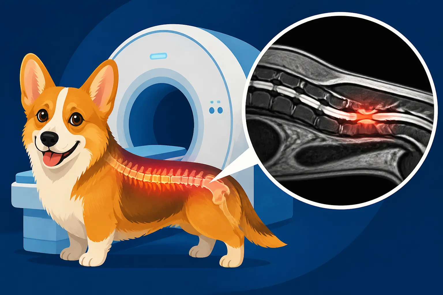
An MRI, or Magnetic Resonance Imaging, creates detailed images of the body using strong magnetic fields and radio waves. For spinal issues, it's considered the gold standard because it can clearly visualize the spinal cord, the intervertebral discs, nerves, and surrounding soft tissues. This allows veterinarians to pinpoint the exact location, nature, and severity of spinal cord compression, which is crucial for planning the most effective treatment. When you're facing potentially serious issues, a comprehensive understanding from an MRI provides the best chance for recovery. It's often an expensive procedure, but reputable pet insurance providers like Healthy Paws can make it more accessible when you need it most.
What an MRI Can Detect Specifically
An MRI gives your veterinary neurologist or surgeon a literal roadmap of your Corgi's spine. Here's what they'll be looking for:
- Disc Herniation (IVDD): This is the big one. An MRI will show exactly which disc (or discs) has ruptured or bulged, the extent to which the disc material has entered the spinal canal, and how much it's compressing the spinal cord. It can differentiate between a sudden extrusion (Type I) and a more gradual protrusion (Type II), which can influence treatment decisions. You’ll get a clear visual of Disc Disease in a Long-Backed Corgi.
- Spinal Cord Edema or Swelling: The spinal cord itself can become swollen or inflamed due to compression. An MRI can detect this edema, indicating the severity of the injury and the potential for neurological damage.
- Spinal Cord Bruising or Hemorrhage: In severe cases of acute disc rupture, there can be bruising or even bleeding within or around the spinal cord. An MRI is excellent at detecting these internal injuries.
- Tumors or Cysts: While less common than IVDD, spinal tumors or cysts can also cause compression. An MRI can identify these growths, distinguishing them from disc material. This is crucial for prognosis and treatment options, as tumors require a very different approach than IVDD.
- Inflammation or Infection: An MRI can also pick up signs of inflammation within the spinal column, such as discospondylitis (inflammation of a disc and adjacent vertebrae), or other infections that might be affecting the spine.
- Vertebral Abnormalities: Although less common, congenital abnormalities in the vertebrae that could contribute to spinal instability or compression can sometimes be seen on an MRI, alongside X-ray findings.
When Your Vet Might Recommend an MRI
Your veterinarian will likely recommend an MRI if your Corgi presents with acute or rapidly worsening neurological signs such as sudden weakness in the hind legs, knuckling over (dragging their paws), inability to stand, severe pain, or loss of bladder/bowel control. These are often considered Emergency Signs of a Corgi Disc Injury. If conservative management (such as strict Crate Rest for a Corgi With a Bad Back and medication) isn't yielding improvement, or if the symptoms are severe from the outset, an MRI becomes a vital step.
It's also highly recommended when surgery is being considered. A surgeon needs a precise map of the affected area to plan the procedure, minimize surgical time, and maximize the chances of a successful outcome. Without an MRI, surgery would be a much riskier, less targeted endeavor. Even in cases of chronic, slowly progressive issues, an MRI can help differentiate between various causes. Knowing How Fast a Corgi Disc Injury Needs Treating often hinges on accurate diagnostics.
Interpreting MRI Results for Your Corgi
Once the MRI is complete, the images are sent to a veterinary radiologist or neurologist for interpretation. This is where all those detailed slices of your Corgi’s spine are meticulously analyzed to form a diagnosis.
Understanding Disc Herniations and Protrusions
When the radiologist looks at the MRI images, they're typically looking for specific changes in the intervertebral discs. In a healthy disc, the nucleus pulposus appears bright on certain MRI sequences due to its water content. In a degenerated disc, it often appears darker, indicating dehydration and hardening. If the disc has herniated (extruded), you'll see the disc material outside its normal boundaries, pressing on the spinal cord. The degree of compression is critical – a little bit of pressure might cause mild pain, while severe compression can lead to paralysis.
The images will also show whether it's an acute extrusion, where a chunk of disc material has burst out, or a chronic protrusion, where the disc has slowly bulged. This distinction helps guide treatment. For instance, a sudden, severe extrusion often necessitates urgent Spinal Surgery on a Corgi, while a mild protrusion might respond well to conservative management like Crate Rest for a Corgi With a Bad Back. The exact location of the herniation also matters immensely; a disc problem in the neck (cervical spine) will present differently than one in the mid-back (thoracolumbar spine), affecting which legs are impacted.
Identifying Other Spinal Cord Compressions
Beyond typical disc issues, an MRI is excellent at spotting other sources of spinal cord compression. Sometimes, inflammation or scar tissue from previous injuries can cause ongoing problems. In older Corgis, a condition called degenerative lumbosacral stenosis might develop, affecting the very end of the spine where it meets the pelvis, leading to hind leg weakness and pain. An MRI can clearly visualize the nerve roots and spinal cord in this area, showing any compression.
Tumors, as mentioned, are another concern. They can originate from the bone, the spinal cord itself, or the surrounding tissues. An MRI can often differentiate tumor tissue from normal tissue and show its exact size and location, which is vital for biopsy planning or surgical removal. Without the detailed images provided by an MRI, these conditions could easily be misdiagnosed or missed entirely, leading to ineffective treatment and prolonged suffering for your Corgi.
Distinguishing Between Conditions Like DM and IVDD
This is one of the most vital roles an MRI plays in a Corgi, especially as they enter The Corgi Years When the Back End Gives Out. Both Degenerative Myelopathy (DM) and IVDD can cause hind leg weakness, ataxia (uncoordinated gait), and even paralysis. However, their treatments and prognoses are vastly different. IVDD is often treatable, especially with surgery, while DM is a progressive, incurable neurological disease.
An MRI of a Corgi with DM will typically show a relatively normal-looking spine, with little to no significant spinal cord compression that would explain the severe neurological deficits. This absence of structural compression is a key indicator that another neurological condition, such as DM, is at play. Conversely, an MRI for IVDD will clearly show disc material pressing on the spinal cord. This is how a neurologist, whom you might consult to know What a Neurologist Checks in a Corgi, uses the MRI to help definitively Tell Disc Pain From DM in a Corgi, even if DM can only be fully confirmed post-mortem. This distinction is crucial for managing your Corgi's care and making informed decisions.
Preparing Your Corgi for an MRI and What to Expect
The idea of your Corgi undergoing an MRI can be daunting, particularly because it requires anesthesia. However, understanding the process can help alleviate some of that natural anxiety.
The Anesthesia Process for a Corgi
Because an MRI machine requires the patient to remain perfectly still for an extended period (often 30-90 minutes, depending on the area being scanned and the number of sequences required), general anesthesia is absolutely necessary for dogs. Before anesthesia, your Corgi will undergo a thorough pre-anesthetic workup. This typically includes blood tests to check organ function, an electrocardiogram (ECG) to assess heart health, and a physical exam. These steps are crucial to ensure your Corgi is a good candidate for anesthesia and to tailor the anesthetic protocol to their individual needs.
During the procedure, your Corgi will be intubated (a breathing tube placed in their windpipe) and carefully monitored by a veterinary technician or anesthesiologist. They will track vital signs like heart rate, breathing, blood pressure, oxygen saturation, and temperature. Anesthesia is always a risk, but with modern protocols and vigilant monitoring, it is generally very safe, even for a breed like the Corgi. Many pet insurance plans, like those from Healthy Paws, often help cover the costs of these diagnostics, including anesthesia, which can be a significant relief during a stressful time.
Post-MRI Care and Next Steps
Once the MRI is complete, your Corgi will be carefully woken up from anesthesia. They'll be monitored closely during recovery, and depending on their condition before the MRI, they might need to stay at the veterinary hospital for a few hours or even overnight. It's not uncommon for them to be a little groggy, disoriented, or even nauseous as the anesthesia wears off.
Once home, you'll receive specific instructions from your vet regarding feeding, medication, and activity restrictions. This might include continued Crate Rest for a Corgi With a Bad Back if the diagnosis indicates it. The most important step, however, is the consultation with your vet or neurologist to discuss the MRI findings. They will explain exactly what the images showed, provide a definitive diagnosis, and outline the recommended treatment plan, whether it's conservative management with medication and rest, or surgical intervention. This is where you'll learn Whether a Corgi Needs Surgery or Rest based on the precise information the MRI provides. They might also discuss options like Physiotherapy for a Corgi Losing Its Legs as part of a comprehensive recovery strategy.
Disclaimer
The information provided in this article is for general informational purposes only and is not intended as a substitute for professional veterinary advice, diagnosis, or treatment. Always seek the advice of your veterinarian or other qualified animal health provider with any questions you may have regarding a medical condition or the health and welfare of your pet.