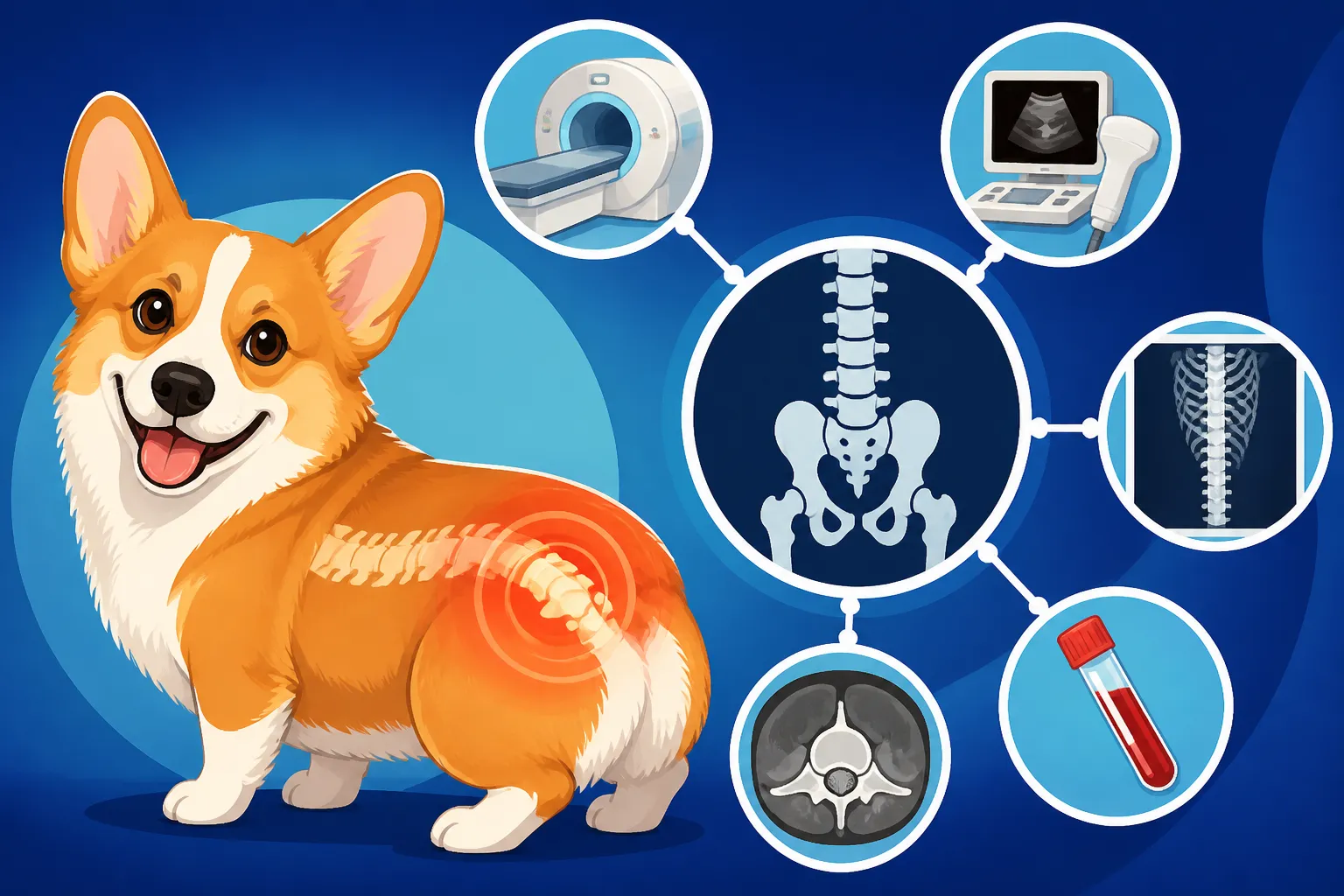
Scans a Corgi Needs Before Back Surgery
Few things are as heart-stopping for a Corgi owner as seeing their beloved companion suddenly struggle with their back legs, perhaps stumbling, crying out, or even dragging a limb. It’s a frightening moment that immediately conjures fears of spinal injury or worse, and the daunting prospect of expensive veterinary care and potentially even surgery. Navigating the world of diagnostic imaging to figure out exactly what’s going on can feel overwhelming, especially when you're worried sick. This guide aims to demystify the essential scans your Corgi might need before back surgery, explaining what each reveals, why it's necessary, and what to expect during this incredibly stressful period.
Why Diagnostic Scans Are Critical for Corgi Back Pain
When your Corgi shows signs of back pain, weakness, or neurological deficits, the first thought that often comes to mind is a slipped disc. While intervertebral disc disease (IVDD) is unfortunately common in breeds with long backs, like our Corgis, it's crucial to remember that a range of other conditions can present with similar symptoms. Without precise diagnostic imaging, your veterinarian is essentially trying to solve a puzzle with half the pieces missing, which can lead to delayed or even incorrect treatment.
Pinpointing the Exact Problem in a Corgi's Spine
The general symptoms of a Corgi struggling with its back—a reluctance to jump, a yelp when lifted, an unsteady gait, or even paralysis—can be indicative of several underlying issues. It could be something as acute as a sudden disc herniation, or a more insidious, progressive condition. Without knowing the exact cause and location of the problem, any intervention, especially surgery, becomes a risky guess. Imagine a surgeon trying to fix a complex problem deep inside the body without a detailed map. That's essentially what happens without proper scans. These sophisticated images provide that map, allowing your vet to see precisely what's happening within your Corgi's intricate spinal column. They illuminate whether the issue is a disc, a bone spur, a tumor, or even an inflammatory condition.
Guiding Surgical Planning and Prognosis for Spinal Injuries
Once a problem is identified, these diagnostic scans become the blueprint for surgical intervention. For instance, if your Corgi has a ruptured disc, the scans will show the exact disc space involved, the direction the disc material has extruded, and the extent of spinal cord compression. This information is absolutely vital. A surgeon needs to know where to operate, how aggressive to be, and what challenges they might encounter. Without this clarity, a surgical approach could be less effective, or worse, lead to complications. The scans also play a significant role in determining the prognosis. The severity of spinal cord compression or the presence of specific types of lesions can help your veterinary neurologist give you a more accurate expectation for recovery, helping you make informed decisions about your Corgi's future.
Differentiating IVDD from Other Conditions
One of the most pressing reasons for advanced imaging is to differentiate IVDD from other neurological disorders that can mimic its signs. For example, Degenerative Myelopathy and the Corgi is a progressive neurological disease that affects the spinal cord, leading to weakness and incoordination in the hind limbs, similar to many disc issues. However, the treatment for DM is entirely different from IVDD surgery, as DM is not a surgically correctable condition. Imaging, particularly MRI, allows vets to clearly distinguish between a physical compression that can be relieved surgically (like a slipped disc) and an internal degeneration of the spinal cord (like DM). This distinction is paramount because proceeding with surgery for DM would be futile and expose your Corgi to unnecessary risks and expense. Conversely, delaying surgery for a severe disc compression can lead to irreversible neurological damage. Understanding Telling a Corgi's Slipped Disc From Myelopathy quickly is critical for the best outcome.
Understanding X-rays and Their Limitations
When your Corgi first exhibits signs of back trouble, routine X-rays are almost always the first diagnostic step your veterinarian will take. They are readily available, relatively inexpensive, and can provide some initial, important clues. However, it's equally important to understand what X-rays can't tell us, especially when it comes to the complex soft tissues of the spine.
What Standard Radiographs Can Reveal
Standard radiographs, or X-rays, provide a two-dimensional image of your Corgi's bone structures. They are excellent for identifying obvious issues like fractures, severe spinal misalignment, or certain types of bony tumors. In the context of IVDD, X-rays might show secondary signs such as narrowed disc spaces between vertebrae, indicating that a disc has lost height, or the presence of calcified disc material within the spinal canal. Calcification is a common precursor to disc herniation, so seeing it on an X-ray can raise a red flag. Your vet might take multiple views—from the side and from above—to get a comprehensive look at the vertebral column. These initial images can help rule out some causes of pain or suggest where further investigation might be needed.
Why X-rays Aren't Enough for Soft Tissue Issues
Here's the critical limitation: X-rays are fantastic for bones, but they are very poor at visualizing soft tissues. This means that a Corgi's slipped disc itself, which is made of cartilage, ligaments, and fluid, will not directly appear on a standard X-ray. You can't see the actual disc material pushing onto the spinal cord. While an X-ray might show a narrowed disc space, it doesn't confirm that the disc has ruptured or, more importantly, where that ruptured material is pressing on the delicate spinal cord. This is a huge problem because the exact location and severity of spinal cord compression are the most crucial pieces of information needed for surgical planning. Relying solely on X-rays for disc disease can lead to an incorrect diagnosis, or even worse, surgery in the wrong spot, which is a devastating outcome for both you and your Corgi.
Myelography: An Older, Invasive Option
Before the widespread availability of advanced imaging like MRI and CT, veterinarians sometimes resorted to a procedure called myelography to get a better look at the spinal cord. This technique involves injecting a special contrast dye directly into the spinal canal, around the spinal cord, while the dog is under general anesthesia. The dye then shows up on X-rays, outlining the spinal cord and revealing any areas where it might be compressed or pinched by disc material or other lesions. While myelography can certainly identify spinal cord compression, it is an invasive procedure with inherent risks. There's a chance of neurological worsening, and in some cases, it can trigger seizures post-procedure. Because of these risks and the superior detail offered by MRI and CT, myelography is now largely considered a last resort or is used in conjunction with CT scans in facilities where MRI is not available.
The Indispensable Role of MRI for Corgi Spines
For any suspected soft tissue injury or compression of the spinal cord, especially those involving intervertebral discs, Magnetic Resonance Imaging (MRI) is widely considered the gold standard in veterinary medicine. It provides an unparalleled level of detail that traditional X-rays simply cannot achieve.
How MRI Provides Unparalleled Detail
MRI utilizes powerful magnetic fields and radio waves, rather than ionizing radiation, to create highly detailed images of soft tissues within the body. When it comes to your Corgi's spine, this means we can see the intervertebral discs themselves, the spinal cord, the surrounding nerves, and even subtle inflammation. An MRI can pinpoint the exact location of a disc herniation, whether it's extruded (material has burst out) or protruded (bulging), the direction of the herniation, and most critically, the precise degree of spinal cord compression. This detailed view allows veterinary neurologists to confidently determine if surgery is needed and, if so, exactly where to operate. For differentiating between a physical disc problem and conditions like degenerative myelopathy, MRI is invaluable. It provides clear answers to questions that clinical examination and X-rays can only hint at, like discerning between Telling a Corgi's Slipped Disc From Myelopathy. The images are so precise that a veterinary radiologist can often predict the best surgical approach and estimate the chances of a full recovery, giving you a much clearer picture of your Corgi's future. This level of insight is crucial for cases where What an MRI Shows in a Corgi Back will define the entire treatment plan.
Preparing Your Corgi for an MRI Scan
Undergoing an MRI is not a quick, in-and-out procedure for pets. Because the machine requires absolute stillness to produce clear images, your Corgi will need to be under general anesthesia for the entire duration of the scan, which can range from 45 minutes to a couple of hours. This means a thorough pre-anesthetic workup is essential. Your vet will typically recommend blood tests to assess organ function and ensure your Corgi can safely metabolize anesthetic drugs. For older Corgis or those with known health issues, additional cardiac screening might be performed.
On the day of the scan, your Corgi will usually be admitted to the veterinary hospital in the morning, having fasted since the night before. The veterinary team will place an intravenous catheter for medication administration and continuously monitor vital signs throughout the anesthesia. While under anesthesia, your Corgi will be carefully positioned within the MRI machine, which can be quite noisy. Rest assured, the veterinary team will be dedicated to keeping your Corgi safe and comfortable throughout the process, focusing on minimizing risks, especially for breeds known for specific health concerns. This often includes performing Clotting Tests Before a Corgi Goes Under to avoid complications.
Cost and Accessibility of MRI Scans
There's no sugarcoating it: an MRI for your Corgi is a significant financial investment. The cost can vary widely depending on your geographic location, the specific veterinary specialty clinic you visit, and the extent of the scan, but you can generally expect to pay anywhere from $2,500 to $5,000 or even more. This price typically includes the anesthesia, the scan itself, interpretation by a board-certified veterinary radiologist or neurologist, and a consultation to discuss the findings.
Many owners find themselves reeling from these unexpected costs, which is why having pet insurance can be an absolute lifesaver. Providers like Healthy Paws offer coverage for unexpected illnesses and accidents, which can certainly include spinal issues requiring MRI and surgery. If you're considering a Corgi, or already have one, looking into comprehensive pet insurance early on is a wise decision to protect against these high-cost emergencies. Unfortunately, MRI machines are not standard equipment in every general veterinary practice. Most require a referral to a specialty veterinary hospital, often those with dedicated neurology departments, which might mean traveling to a larger city or a university veterinary teaching hospital.
When a CT Scan Comes into Play
While MRI is the go-to for soft tissue details, there are specific situations where a Computed Tomography (CT) scan is either preferred or provides complementary information for diagnosing your Corgi's spinal problem. It's another powerful tool in the diagnostic arsenal, offering a different perspective on the intricate structures of the spine.
CT vs. MRI: Different Strengths
CT scans use a series of X-ray images taken from various angles around your Corgi's body, which are then processed by a computer to create cross-sectional (slice-like) images. While both CT and MRI can produce detailed images, they excel in different areas. CT scans are superior for visualizing bone structures with incredible clarity. This means if your vet suspects a fracture, a bony tumor, or issues related to spinal instability involving the vertebrae themselves, a CT scan often provides clearer detail than an MRI. It's also much faster than an MRI, which can be a critical factor in emergency situations where rapid diagnosis is paramount. However, CT scans are generally less effective at distinguishing between different types of soft tissues, like the nuances between a healthy disc and a herniated one, or the precise extent of damage to the spinal cord, compared to MRI.
Specific Scenarios for CT Recommendation
So, when might a CT scan be recommended for your Corgi? One common scenario is in cases of suspected trauma, such as after a fall or an accident, where a spinal fracture or severe bony injury needs to be quickly identified. CT's speed and excellent bone detail make it ideal for these urgent situations. It's also often used if an MRI is unavailable or contraindicated for some reason. In such cases, a CT scan might be performed in conjunction with myelography (injecting contrast dye into the spinal canal), as the dye can help highlight areas of spinal cord compression that wouldn't be as clear on a standalone CT. This combined approach tries to bridge the gap in soft tissue imaging that CT alone has. Furthermore, some veterinary specialists prefer CT for pre-surgical planning in certain complex cases, particularly if the problem has a significant bony component.
Anesthesia and Costs for CT Scans
Just like with an MRI, a CT scan requires your Corgi to be perfectly still for accurate imaging. This means general anesthesia is necessary. The preparation is very similar: pre-anesthetic blood work, fasting, and continuous monitoring of vital signs throughout the procedure. The good news is that because CT scans are generally much faster than MRIs, the anesthesia time is often shorter, which can sometimes reduce the overall risk.
In terms of cost, CT scans are typically less expensive than MRIs but still represent a substantial expense. You can expect a CT scan to range from approximately $1,500 to $3,500, depending on the clinic and region. Again, pet insurance can be incredibly helpful in mitigating these costs, making it easier to afford the best diagnostic care for your Corgi when spinal issues arise. Always have a thorough discussion with your veterinarian about which imaging modality is best suited for your Corgi's specific condition, weighing the diagnostic benefits against the costs and risks of each option. This discussion should cover How Fast a Corgi Disc Injury Needs Treating and what the most urgent next steps are.
Interpreting the Results and Making Decisions
Once the scans are complete, the real work of interpretation begins. This critical phase involves specialized veterinary professionals and ultimately leads to the treatment plan for your Corgi's back issue. Understanding this process will help you feel more empowered and less adrift during an anxious time.
Collaborative Review with Your Veterinary Neurologist
The detailed images from an MRI or CT scan are not typically interpreted by your general practice veterinarian alone. Instead, they are sent to a board-certified veterinary radiologist or, more commonly for spinal issues, a veterinary neurologist. These specialists have extensive training and experience in reading complex imaging studies and can accurately identify subtle abnormalities that might be missed by a less specialized eye. They will generate a comprehensive report outlining their findings, the presumed diagnosis, and any recommendations. This report is then shared with your primary veterinarian, and most importantly, with you. It's crucial during this consultation to ask every question you have. Don't hesitate to ask for explanations of medical terms, to see the images yourself, and to fully understand what has been found. This collaborative approach ensures that the best minds are looking at your Corgi's case and that you, as the owner, are fully informed.
Weighing Surgical vs. Conservative Management
With a clear diagnosis in hand from the imaging, the discussion then shifts to treatment options. This is often the most difficult part for owners. The decision between surgical intervention and conservative (non-surgical) management is complex and depends on several factors: the severity of the spinal cord compression, the type of injury (e.g., acute rupture vs. chronic degeneration), the duration of symptoms, your Corgi's overall health, and your financial capabilities.
For severe disc herniations or rapidly progressing neurological deficits, surgery is often the recommended course of action. This is particularly true if your Corgi has lost the ability to walk or has significant pain and weakness. The goal of surgery is to decompress the spinal cord, removing the offending disc material or bony spurs, to alleviate pressure and allow the nerves to recover. If the scans indicate a severe injury, the question of Whether a Corgi Needs Surgery or Rest becomes less a matter of choice and more a matter of urgency. However, for milder cases, or if surgery isn't a viable option due to financial constraints or other health concerns, conservative management might be attempted. This typically involves strict crate rest for several weeks (often 4-6 weeks), pain medication, anti-inflammatory drugs, and muscle relaxants. It’s a challenging path, requiring immense dedication to keep an active Corgi confined, but it can be effective for some. If you go this route, you'll want to ensure you have all the necessary supplies for comfortable crate rest, from orthopedic beds to puzzle toys, which can be found readily on sites like Chewy.
Prognosis and What to Expect Post-Diagnosis
The prognosis for your Corgi after a spinal injury, whether treated surgically or conservatively, is heavily influenced by what the diagnostic scans reveal. Factors like the degree of spinal cord compression, how long the compression has been present, and the overall health of the spinal cord tissues (seen on MRI) all play a role. Your neurologist will use this information to give you a realistic outlook on recovery.
Recovery can be a long road, often involving weeks or even months of careful rehabilitation. This might include services like hydrotherapy, physiotherapy, or even acupuncture, especially for Corgis who have lost significant function in their hind legs. The path to recovery for Recovery Rates After Corgi Disc Surgery is not always straightforward, and patience is key. Ongoing management, including weight control (Why Corgi Weight Is a Spinal Problem is a real concern), appropriate exercise, and perhaps even joint supplements, will be crucial to prevent future episodes. Even after recovery, some Corgis may have residual neurological deficits. The goal is always to restore the best possible quality of life for your furry friend, ensuring they can enjoy their "Corgi years" to the fullest. Offering a high-quality, balanced diet, perhaps even fresh-prepared options like those from Ollie, can also support overall health and recovery during this time. Remember that The Corgi Years When the Back End Gives Out can be tough, but proper care can make a huge difference.
Comparison of Diagnostic Scans for Corgi Spinal Issues
To help clarify the differences and applications of the various diagnostic scans, here’s a quick overview:
| Scan Type | What it Shows Best | Pros | Cons | Typical Cost Range (USD) |
|---|---|---|---|---|
| X-ray | Bone structure, alignment, obvious fractures, narrowed disc spaces | Widely available, relatively inexpensive, quick | Poor soft tissue detail, doesn't show discs or spinal cord directly | $150 – $400 |
| Myelography | Spinal cord compression (indirectly via dye) | Better than X-ray for compression, still used if MRI/CT unavailable | Invasive, risk of neurological worsening/seizures, superseded by MRI | $800 – $1,500 |
| CT Scan | Bone detail, fractures, bony tumors, rapid emergencies | Faster than MRI, excellent bone detail, 3D views | Less soft tissue detail than MRI, radiation exposure | $1,500 – $3,500 |
| MRI Scan | Soft tissues (discs, spinal cord, nerves), inflammation, exact type/severity of disc disease | Gold standard for disc disease, unparalleled soft tissue detail | Expensive, longer procedure, requires specialized equipment/staff | $2,500 – $5,000+ |
Disclaimer
The information provided in this guide is intended for educational purposes only and should not be considered a substitute for professional veterinary advice. Always consult with a qualified veterinarian or veterinary specialist for any health concerns or before making any decisions about your Corgi's medical care.